Two Valves Repaired: Seeing the Heart Work Better
One of the things I love most about cardiac surgery is the ability to see a serious problem and, just a few hours later, see the heart working dramatically better.
With permission from the patient to share this case, I want to show you an example of that transformation.
This 50-year-old patient came to us with severe mitral regurgitation caused by mitral valve prolapse, along with significant tricuspid valve regurgitation.
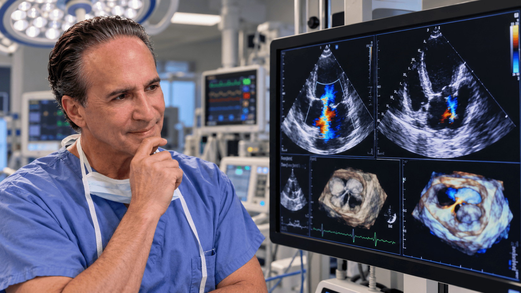
The video accompanying this post shows the intraoperative transesophageal echocardiogram (TEE) before and after surgery. We were able to successfully repair both valves, preserving the patient’s own valves rather than replacing them.
What Was Wrong With the Mitral Valve?
The mitral valve sits between the left atrium and left ventricle. It allows blood to flow forward into the ventricle and then closes to prevent blood from flowing backward.
In this case, part of the posterior mitral valve leaflet was prolapsing because of a torn chord. These supporting structures help the valve leaflets close properly.
When the leaflet prolapses, the valve cannot close completely. Blood leaks backward into the left atrium, a condition called mitral regurgitation.
The preoperative echocardiogram showed a significant regurgitant jet, confirming that the valve was leaking severely.
The Tricuspid Valve Was Leaking, Too
The patient also had severe tricuspid regurgitation.
The tricuspid valve sits between the right atrium and right ventricle. In this case, the valve was leaking primarily because the valve annulus, the ring-like structure supporting the valve, had become dilated.
The annulus measured 43 millimeters. When the annulus becomes enlarged, the valve leaflets may no longer come together properly, allowing blood to leak backward.
This required a repair of the tricuspid valve as well.
Repairing the Mitral Valve
Rather than replacing the mitral valve, we were able to repair the patient’s own valve.
We placed four Gore-Tex chords in the posterior leaflet to correct the prolapse caused by the torn chord. These artificial supporting structures help restore the normal position and function of the valve leaflet.
We also used an annuloplasty ring to support the valve and help maintain its shape.
After the repair, the echocardiogram showed good leaflet coaptation, meaning the valve leaflets were coming together properly.
There was no further mitral regurgitation. The patient also had no systolic anterior motion, or SAM, and the measured gradient across the valve was only 2.
Repairing the Tricuspid Valve
The tricuspid valve was repaired with an annuloplasty ring to address the dilated annulus.
The goal was to restore the valve’s ability to close properly and prevent blood from leaking backward.
After the repair, the echocardiogram showed no further tricuspid regurgitation.
Checking the Heart After Repair
Intraoperative echocardiography allows us to evaluate the heart immediately after surgery.
We checked both repaired valves and confirmed that they were functioning well. We also evaluated the aortic valve to make sure that the mitral and tricuspid repairs had not distorted its leaflets or caused aortic insufficiency.
The aortic valve looked good, with no aortic insufficiency.
Both valve repairs were successful. The patient had good biventricular function and was in normal sinus rhythm.
Why Repair the Patient’s Own Valves?
When appropriate, repairing the patient’s own valve can restore more normal blood flow while preserving the natural valve.
Severe valve regurgitation can make the heart work harder. Over time, that extra workload can affect the heart’s ability to function efficiently.
When a valve can be repaired successfully, we can correct the leak while preserving the patient’s own anatomy.
That is why valve repair is often preferred when it is technically feasible and appropriate for the patient.
The Left Atrial Appendage
We also closed the left atrial appendage during this operation.
The left atrial appendage is a small pouch connected to the left atrium. In some patients, particularly those who develop atrial fibrillation, blood can collect there and form a clot.
Closing the appendage can help reduce the risk of stroke should atrial fibrillation develop in the future. The decision to close it depends on the patient’s individual circumstances.
The Difference a Repair Can Make
For me, there is still something extraordinary about watching a severely leaking valve become competent again and seeing the immediate difference on the operating-room echo.
At the beginning of the operation, we could see the abnormal leaflet motion and the significant regurgitant jets.
After the repairs, the valves were closing properly, the regurgitation was gone, and the heart was functioning well.
That is the value of intraoperative echocardiography. It allows us to see the problem, guide the repair, and confirm the result before the patient leaves the operating room.
Simple educational takeaway: When a heart valve leaks severely, the heart has to work harder. When appropriate, repairing the patient’s own valve can restore more normal blood flow while preserving the natural valve.
Repair when we can. Preserve the patient’s own heart whenever possible. ❤️
Interested in my book? You can buy it on Amazon!
(𝘛𝘩𝘦 𝘷𝘪𝘦𝘸𝘴 𝘦𝘹𝘱𝘳𝘦𝘴𝘴𝘦𝘥 𝘪𝘯 𝘮𝘺 𝘱𝘰𝘴𝘵𝘴 𝘢𝘳𝘦 𝘮𝘺 𝘰𝘸𝘯 𝘢𝘯𝘥 𝘥𝘰 𝘯𝘰𝘵 𝘳𝘦𝘱𝘳𝘦𝘴𝘦𝘯𝘵 𝘵𝘩𝘦 𝘷𝘪𝘦𝘸𝘴 𝘰𝘧 𝘮𝘺 𝘦𝘮𝘱𝘭𝘰𝘺𝘦𝘳 𝘰𝘳 𝘢𝘯𝘺 𝘰𝘳𝘨𝘢𝘯𝘪𝘻𝘢𝘵𝘪𝘰𝘯.)
Recent Posts

It’s Never Too Late to Invest in Your Health
You don’t need to be an athlete or 25 to invest in your health. Discover why movement, emotional well-being, and purpose matter at every age.

73 Years Young: Strength Has No Age Limit
At 73, Joseph Lisa is still getting stronger. Discover why age doesn’t have to define our limits and why staying active matters for a longer, healthier life.

Should We Be Afraid of AI?
Should we be afraid of AI? Dr. Scott Silverstein, a pioneer in medical informatics, shares why human judgment, experience, and responsible innovation matter more than ever.

Extraordinary Care Around the Clock
While most of us are sleeping, the cardiac surgery night crew at Jefferson Einstein Montgomery Hospital is working tirelessly to keep patients safe, supported, and cared for. Their dedication, compassion, and teamwork make extraordinary care possible around the clock.

Not Every Patient With Aortic Stenosis Is Best Served by the Least Invasive Approach
Not every patient with aortic stenosis is best served by the least invasive approach. For this 68-year-old patient, severe aortic stenosis and critical coronary artery disease made surgical valve replacement and CABG the right choice.

When the Right Heart Valve Changes: A Fourth Operation After 14 Years
A patient’s remarkable valve history led to a fourth open-heart operation, not because her mechanical valve failed, but because her medical circumstances changed.
Featured
Pages
- Learn about heart valves. Heart Valves
- Read testimonials. Testimonials
- Did you know I have a consulting firm? Singer Heart/Lung Consulting
- Check out my TedTalk! Defining Success
Links
- Links page with more information about your heart. Links
- Dr. Adam Pick's Site: heart-valve-surgery.com