The tricuspid valve lies between the right atrium and the right ventricle. Blood returning to the heart from the body will enter the right atrium and pass through the tricuspid valve into the right ventricle and then on to the lungs.
Similar to the pulmonary valve, it is unusual to primarily operate upon an adult’s tricuspid valve.
The two most common reasons to operate on an adult’s tricuspid valve are:
- So-called “Functional” Tricuspid Regurgitation
- Infection of the Tricuspid Valve
Functional Tricuspid Regurgitation
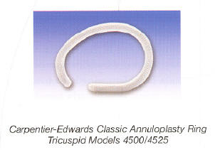
Tricuspid Ring
When patients have high pressures in their lungs, usually from mitral valve disease, poor left ventricular function, or primary lung disease, the pressure on the right ventricle could be so great that the right ventricle enlarges rendering the tricuspid valve incompetent.
To reverse this condition, usually the surgeon only needs to place a prosthetic ring around the tricuspid valve so as to return it to it’s normal size. It almost never needs to be replaced for this condition.
Infection of the Tricuspid Valve
Endocarditis (infection) of the tricuspid valve is rare.
Unfortunately, it usually occurs in patients who are intravenous drug users. By using dirty needles, the blood returning to the heart is contaminated resulting in terrible infections of the tricuspid valve.
This is the one condition that usually requires replacing the tricuspid valve with a prosthetic valve, usually a porcine (pig) stented valve as shown below.
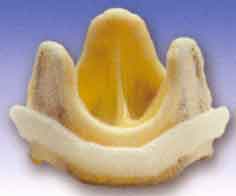
Porcine (pig) Stented Valve
Case Study
Below you can see a case of combined mitral valve and tricuspid valve repair. This video explains the anatomy as seen on trans esophageal echocardiogram (TEE) and my techniques for repair in the operating room.
TEE using both 2D and 3D imaging has become an invaluable tool to successfully perform mitral and tricuspid valve repair surgeries. The use of intraoperative TEE is a collaborative effort by the cardiac surgeon, cardiologist, and cardiac anesthesiologist.
Below is a brief video showing a combined mitral and tricuspid repair, illustrating the importance of having a team that is experienced with intraoperative TEE 2D and 3D anatomy.