Most people don’t realize that there can be tumors of the heart. In fact, tumors of the heart are not uncommon.
Heart tumors can be either benign or malignant. Approximately 70% of primary heart tumors are benign and 30% are malignant. Unfortunately, the heart can also be the site of metastatic tumors from other sites.
Benign Heart Tumors (70%)
- Myxoma
- Lipoma
- Papillary Fibroelastoma
- Rhabdomyoma
- Fibroma
- Hemangioma
- Teratoma
- Mesothelioma of AV Node
- Granular Cell Tumor
- Neurofibroma
- Lymphangioma
Malignant Heart Tumors (30%)
- Angiosarcoma
- Rhabdomyosarcoma
- Mesothelioma
- Fibrosarcoma
- Malignant Lymphoma
- Extraskeletal Osteosarcoma
- Neurogenic Sarcoma
- Malignant Teratoma
- Thymoma
- Leiomyosarcoma
- Liposarcoma
- Synovial Sarcoma
Atrial Myxoma
Atrial myxomas are tumors that occur within the chambers of the heart, usually the left atrium. They account for 29% of all heart tumors. Although generally considered benign, there are reports of metastasis to other parts of the body.
80-90% of myxomas occur in the left atrium. They are usually 5-6 cm in size, but can range from 1-15 cm. Left atrial myxomas are usually attached to the septum of the heart separating the left and right atrium.
Their appearance is typically that of a gelatinous ball projecting into the left atrial chamber, with soft projections, sort of like a small jellyfish! When removing one of these tumors, it is important not to have any of this soft material break off and drop into the heart.
Since these tumors are usually attached the the septal lining, a combination of a left atrial and right atrial incision is often required. A portion of the septum between the left and right atrium is removed with the tumor and the septum is patched with a piece of the patient’s own pericardium or a piece of processed bovine (cow) pericardium.
Case Report 1
Below is a typical case of a left atrial myxoma. The tumor was relatively small and was able to be removed with a single incision in the patient’s left atrium.
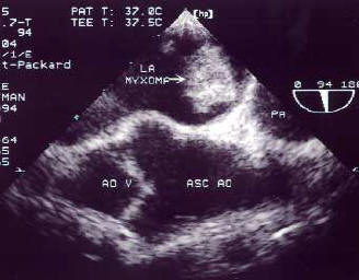
TEE (Trans-Esophageal Echocardiogram) showing a left atrial myxoma
Below is a black and white photograph of a pathologic specimen of the above tumor. Note the jelly-like appearance of the cut tumor.
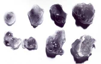
Pathologic Specimen of Tumor
Case Report 2
This case shows an extraordinarily large left atrial tumor. An incision was made in both the left and right atrium and a portion of the atrial septum was removed in order to safely remove the tumor.
 showing a giant left atrial myxoma")
TEE (Trans-Esophageal Echocardiogram) showing a giant left atrial myxoma
Below is a color photograph of a pathologic specimen of the tumor. This tumor was surprisingly solid.

Solid Left Atrial Myxoma Tumor
Here’s a really cool video showing my removing a large left atrial myxoma. These are relatively rare operations, so to see a real video is special.